

Research focus and scientific vision
Research focus and vision
The research group is focused on advanced light microscopy and applications in nanotechnology, cell biology, cancer and neuroscience research.
Advanced light microscopy: Holographic Incoherent Quantitative Phase Imaging (hiQPI)
- novel optical systems (geometric-phase optics)
- novel imaging approaches (3D hiQPI and imaging through turbid-media)
- novel image processing methods (evaluation of dynamics imaged in time series, artificial-intelligence approach)
Vision: to establish hiQPI as a typical technique of advanced light microscopy
Applications of hiQPI in nanotechnology, cell biology and cancer research
- study of phase effects of nanostructured metasurfaces
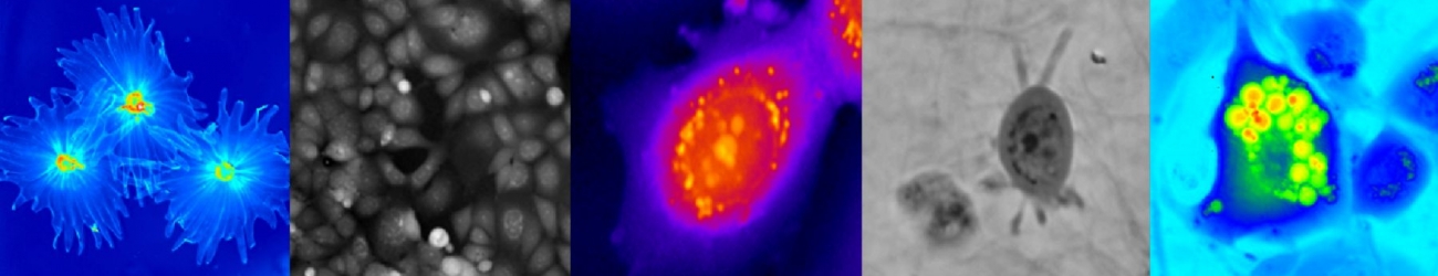
- study of live cancer cell behavior (biopsy-derived cells, intracellular mass dynamics as a biomarker of the malignant cell phenotype)
- strategy for personalized cancer treatment (DANTE: Dynamic Analysis for Neoplasia Treatment with Explants)
Vision: hiQPI assessment of live cancer biopsy cells reactions to therapeutics under consideration as well as preclinical evaluation of potentially new anticancer drugs particularly of migrastatic type
Scientific vision
New dimensions in biomedical microscopy of living cells & tissues are currently emerging worldwide. Maturing holographic microscopy that brought about attributes of quantitative phase imaging (QPI), particularly with low coherence light, as new tools for assessing live cell reactions, is becoming an important part of this process. The RG research and development focuses on fundamental aspects, methods and tools. We have to pay particular attention to applications of this imaging modality in cell biology. This is because of QPI novelty in bringing data on dry mass of cells. This is a parameter, which cell biologists do not count on as indispensable yet. Therefore, our task is to show what new information QPI can reveal about the behaviour of a particular cell phenotype. This endeavour will continue in next years with the emphasis on 3D and turbid-media imaging and finding apt biomedical and clinical applications. Research and development in multimodal holographic microscopy and other innovative methods of advanced light microscopy, such as two-photon microscopy and new generation optical components (such as spatial light modulators and 4G optics) based techniques, will further extend in close connection to the biomedical applications such as cell biology, cancer and neuroscience research. There are unmet needs for biomedical imaging of semi-transparent structures such as fragments of tissue or cells in collagen and we will enhance our focus in the development of QPI techniques for turbid media and 3D imaging. New approaches to QPI imaging will require further development of image processing, dynamic morphometry and cell classification.
An important biomedical project is the development of a strategy for personalised cancer treatment based on live-cell dry-mass profiling, producing a system we call DANTE (Dynamic Analysis for Neoplasia Treatment with Explants). The main objective of this project is the improvement of cancer treatment since cancer-related mortality and morbidity has been increasing worldwide. The growth of cancer cells is not the main problem for cancer patients, the majority of deaths are due to invasion and metastasis. Problems lie in the accuracy of diagnosis and in the choice of the most effective treatment. Currently, the biopsy material is only fixed and investigated by histopathology. We plan to achieve our objective by complementing the standard histopathology with novel pretesting of chemotherapeutic drugs using a time-lapse recording of live biopsy cells. Using part of the biopsy material which is kept alive, may seem obvious. However standard microscopy techniques lack accuracy, speed and are difficult to quantify. Moreover establishing the live cell culture is relatively complicated. This project, therefore, is based on QPI for dry-mass profiling of live cells in tissue culture. Measurements of growth rapidly reveal the mass increase in the individual cells and there is no need to wait for cell divisions. Most importantly, cell motility, essential for invasion and metastasis, can also be rapidly measured in terms of protrusion/ retraction of the cell margin and there is no need to wait for distant translocations. Moreover, the mass profiling microscopy techniques expose the cells to very low phototoxicity. We propose that the potentially effective drug or a combination can be identified by the statistically significant reduction in the rate of motility as well as growth. In the long term, we will be assessing the efficiency of our approach by monitoring the success of the treatment of the donor patients in relationship to our dry-mass profiling data. Our vision is that this approach will lead to a fully automated robotised processing of fresh biopsy material and producing treatment recommendation in a few days.
Collaboration in neuroscience on cellular and molecular aspects of the hemodynamic response of brain will be pursued with the reduced capacity of Hana Uhlířová, whose main contract is currently with the Institute of Scientific Instruments, Czech Academy of Sciences, Brno (Čižmár group).
Completely new field of Biophotonics research has been triggered by collaboration with the Medicem company in the field of visual optics. Multifocal, mainly bifocal and also trifocal, intraocular lenses (IOL) of different designs are now the prevalent solution of cataract and presbyopia correction allowing simultaneous vision mainly at far and near distances. Different strategies can be used to extend the depth of field (DoF), e.g. multifocal IOLs with very low dioptric addition and/or IOL with the controlled amount of aberrations. The main goal of the project is to theoretically and experimentally investigate the impact of these strategies and/or their combinations on DoF and retinal image quality.
The QPI technique is also uniquely suitable for observations and measurements of optical properties and effects of nanotechnology structures such as plasmonic antennas and we envisaged further development and more collaborative applications in this field.
Building the team of solvers composed of all professions like physicists, engineers, statisticians, biologists and medics is essential for the final success. It is the recognition of only recent time that this kind of research team cannot be substituted by only one or two interdisciplinary professionals, who are otherwise vital for core facilities and we try to educate them. In our team, each member should be firmly anchored in his/her specialization and qualified to understand the other views in order to create inclusive plans for solutions to the problems tackled.